Science
Researchers at UVic Revolutionize Microscopy with New Technique
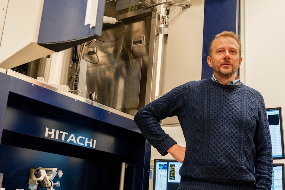
A team of researchers at the University of Victoria has achieved a significant breakthrough in the field of electron microscopy, enabling scientists to visualize atomic-scale structures using more affordable and energy-efficient equipment. Under the leadership of Arthur Blackburn, co-director of UVic’s Advanced Microscopy Facility, this innovation promises to transform microscopy techniques worldwide.
The research, published in Nature Communications, introduces a novel imaging method that allows for sub-Ångström resolution—less than one ten-billionth of a meter—using a compact, low-energy scanning electron microscope (SEM). Previously, such high-resolution imaging was only feasible with large, expensive transmission electron microscopes (TEMs).
“This work shows that high-resolution imaging doesn’t have to rely on expensive, complex equipment,” Blackburn stated. As the Hitachi High-Tech Canada Research Chair in Advanced Electron Microscopy, he emphasized the potential of simpler SEMs when paired with advanced computational techniques. The new approach can achieve resolutions that rival or even surpass traditional methods, making cutting-edge microscopy more accessible to laboratories globally.
The breakthrough utilizes overlapping patterns of scattered electrons to create highly detailed images of samples. This technique enabled the team to achieve a resolution of just 0.67 Ångström, smaller than the size of an atom and approximately one-tenth the width of a human hair.
Implications for Multiple Scientific Fields
The implications of this advancement extend across various scientific disciplines. Blackburn highlighted that this technology could be transformative for fields such as materials science, nanotechnology, and structural biology. “The advance will most immediately benefit the research and production of 2D materials, which are promising in the development of next-generation electronics,” he explained.
In the long term, this technique could also assist in determining the structures of small proteins, offering new avenues for research in health and disease. The accessibility of high-resolution imaging could lead to breakthroughs that enhance our understanding of biological processes and material properties.
As researchers around the world begin to explore the capabilities of this new imaging technique, the potential for innovation and discovery is vast. This achievement not only marks a pivotal moment for the University of Victoria but also sets a new standard for electron microscopy, paving the way for more inclusive scientific exploration.

Japan Elects Sanae Takaichi as Its First Female Prime Minister

Polish Authorities Detain Eight in Sabotage Investigation

Ottawa School Board Allows Meeting Recordings, Bans Live-Streaming

HSBC Appoints David Lindberg as New UK Business Leader

Calgary Mayoral Race Tightens as Farkas Leads Sharp in Unofficial Results

Global Seed Bank Secures Future of Biodiversity for Generations

NovaBay Pharmaceuticals Achieves Compliance with NYSE Standards

Southern Alberta Communities Elect Mayors and Town Councils

Hong Kong Runway Set to Reopen After Cargo Plane Crash

Toyoake City Proposes Daily Two-Hour Smartphone Use Limit

B.C. Review Reveals Urgent Need for Rare-Disease Drug Reforms

Pedestrian Fatally Injured in Esquimalt Collision on August 14

Dark Adventure Game “Bye Sweet Carole” Set for October Release

Jimmy Lai’s Defense Challenges Charges Under National Security Law

Konami Revives Iconic Metal Gear Solid Delta Ahead of Release

Snapmaker U1 Color 3D Printer Redefines Speed and Sustainability

AION Folding Knife: Redefining EDC Design with Premium Materials

Gordon Murray Automotive Unveils S1 LM and Le Mans GTR at Monterey
-
 Science2 months ago
Toyoake City Proposes Daily Two-Hour Smartphone Use Limit
-
 Health2 months ago
B.C. Review Reveals Urgent Need for Rare-Disease Drug Reforms
-
 Top Stories2 months ago
Pedestrian Fatally Injured in Esquimalt Collision on August 14
-
 Technology2 months ago
Dark Adventure Game “Bye Sweet Carole” Set for October Release
-
 World2 months ago
Jimmy Lai’s Defense Challenges Charges Under National Security Law
-
 Technology2 months ago
Konami Revives Iconic Metal Gear Solid Delta Ahead of Release
-
 Technology2 months ago
Snapmaker U1 Color 3D Printer Redefines Speed and Sustainability
-
 Technology2 months ago
AION Folding Knife: Redefining EDC Design with Premium Materials
-
 Business2 months ago
Gordon Murray Automotive Unveils S1 LM and Le Mans GTR at Monterey
-

 Technology2 months ago
Solve Today’s Wordle Challenge: Hints and Answer for August 19
-

 Lifestyle2 months ago
Victoria’s Pop-Up Shop Shines Light on B.C.’s Wolf Cull
-

 Technology2 months ago
Apple Expands Self-Service Repair Program to Canada